
Confidently Identify and Target Lesions¹
Whether using traditional 2D imaging or the latest tomosynthesis (3DTM) imaging, the Affirm® prone system employs advanced, proprietary technology to help clinicians identify, target and access lesions for faster biopsies.1,4 The high-quality imaging of the Affirm® prone biopsy system allows clinicians to pinpoint extremely subtle lesions, including low contrast distortions and faint calcifications, quickly and confidently.¹
Experience the Difference
- Generate high-quality images in seconds
- Identify challenging lesions with a direct-conversion selenium detector and a 14.3 cm x 11.7 cm field of view that is more than 6.5x larger than previous generations of prone biopsy systems1
- Accelerate procedures without manually positioning the X-ray tube1
Identify Challenging Lesions
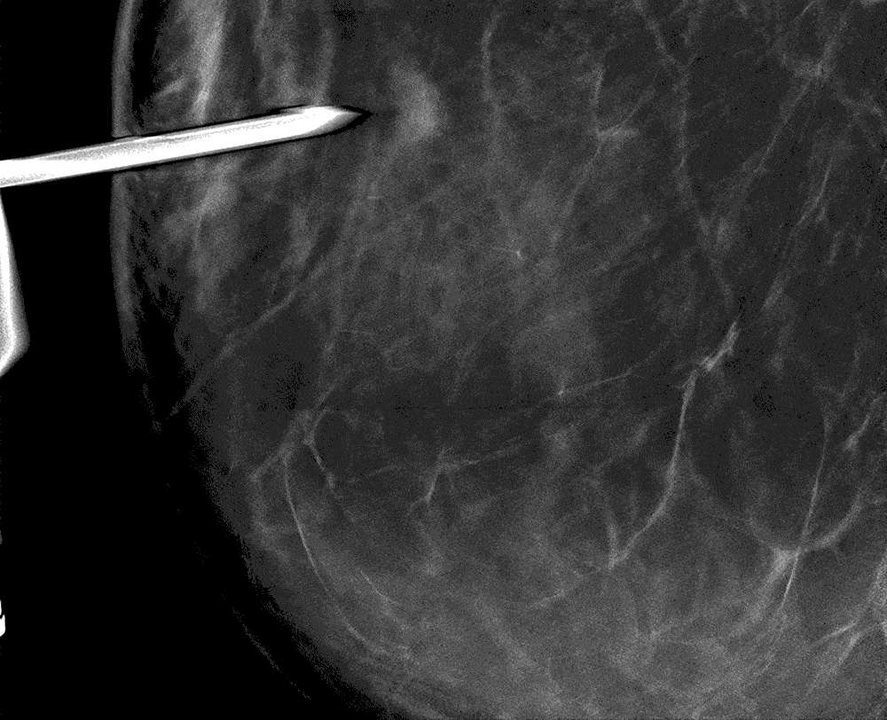
Clinicians can target lesions with confidence using the same powerful imaging technology that drives the Hologic Selenia ® Dimensions® mammography system. The Affirm® prone system’s game-changing capabilities let clinicians see more, helping to pinpoint subtle lesions and faint calcifications that might not be visible with older technology1 – and ultimately may help reduce the number of patients sent to open surgical biopsy.

Broaden the Field View
With a detector that’s more than 6.5 times larger than previous generations of prone biopsy systems, the Affirm® prone system provides an expansive view of the breast to allow for easy targeting. 1,2 The system’s X-ray translucent, clear paddles help visualize tissue inside and outside the biopsy window to quickly and confidently position the patient.

3D™ Biopsy Capable
It’s the only dedicated prone biopsy system offering high-quality 2D or 3D™ imaging, and it utilizes the proven detector technology from Hologic’s Selenia® Dimensions® mammography system.1,4 3D™ Breast Biopsy can localize subtle lesions that may not be visible using conventional 2D imaging.5 It also further reduces process steps, which may lead to a faster procedure and a lower dose of radiation for the patient.2,5 The system’s software is easily upgradable from 2D to 3D™ imaging at any time.

Features
- High resolution direct-conversion amorphous selenium detector.1 The same detector technology found in our Selenia® Dimensions® mammography system.
- More than 6.5 times larger field of view (14.3 cm x 11.7 cm)1
- 3D™ biopsy-capable (including all the capabilities of the 2D configuration)5
- Easily upgradable from 2D to 3D™ imaging
- X-ray translucent, clear paddles
Capabilities and Advantages
Broader Field of View to Identify Challenging Lesions1
X-ray translucent, clear paddles and an expanded, 14.3 cm x 11.7 cm field of view allow visualization of more tissue helping clinicians quickly and easily pinpoint subtle lesions and faint calcifications.1 The larger field of view makes repositioning easy when the lesion is outside of the biopsy window.
Superior Image Quality1
A high resolution, direct-conversion selenium detector provides enhanced 2D and 3D™ imaging.1,2,4
Faster, Efficient Procedures1
A single press of a button automatically moves the tube head over the breast in seconds to acquire all necessary images whether a stereo pair or tomosynthesis sweep.
Clear Guidance
Featuring a 2 MP DICOM compliant monitor as standard with an optional 3 MP monitor upgrade for enhanced image quality and less need to pan and zoom.
Easily Upgradable to 3D™ Breast Biopsy
Visualize Lesions Better1
To fully take advantage of the Affirm® prone biopsy system capabilities, facilities can start with 2D imaging and upgrade to tomosynthesis guided biopsy when ready. This innovative technology helps localize subtle lesions that may not show up with 2D imaging.5
Fewer Images Required5
When using 3D™ technology, the Affirm® prone system further combines steps in the process, which may lead to fewer exposures, a faster procedure, and up to 50% lower radiation dose for patients.5,6


Overall Procedure Dose Comparison (mGy)
| Standard 2D Biopsy Procedure | Standard 3DTM Biopsy Procedure | Full 3DTM Biopsy Procedure | |
|---|---|---|---|
| Scout | 1.6 | 1.8 | 1.8 |
| Targeting | 3.2 | - | - |
| Prefire | 3.2 | 3.2 | 1.8 |
| Postfire | 3.2 | 3.2 | 1.8 |
| Post-procedure | 3.2 | 1.8 | 1.8 |
| Total | 14.4 | 10 | 7.2 |
| % Reduction (Compared to 2D) |
- | 31% | 50% |
Based on dose at 4.2 cm, 50% fatty 50% glandular tissue. Assuming 1.6 mGy per 2D image and 1.8 mGy per tomosynthesis imaging.

“Because of the larger field of view, it's easier and quicker to find lesions."
Hear from Dr. Margot Henebiens about her experience with the Affirm® prone biopsy system.